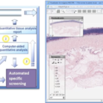
- The imaging tool is a handheld tool, which are two cameras, which take simultaneous clinical photos at from a different angle and when put the scope on patient’s skin lesion.
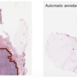
- This gives us skin sections in horizontal planes which go deeper and deeper (from superficial to deep). These images appear on a monitor, like you’re looking at an MRI or x-ray, but in this case, the resolution is very high, like up to four microns, and you can actually see the cells and quickly tell the patient (in experienced hands) is the lesion is benign, malignant or required further investigation.
Advantages
- It’s simple, it’s practical.
- No biopsy is required (which leaves a scar)
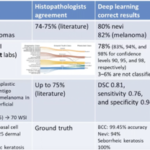
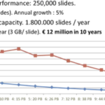
- In teleconfocal these images transferred via the internet to a distant reader (for example to sign the case or a colleague for a second opinion). After looking at the images a diagnosis can quickly be made, a report generated.
Babar Rao, MD. The Emerging Role of Tele-confocal and Tele-pathology in Global Dermatology. 8th World Congress of Teledermatology, Skin Imaging and AI in Skin diseases – November 2020