Digital Dermpath: Deep-Learning On Classic Digital Slides
- Paraffin sections are classically stained with Hematoxylin-Eosin (HE) before being integrated in a glass slide.
- One step further takes us to the digital slide.
These studies below highlight how deep learning algorithm development has been demonstrating its usefulness on H&E stained skin biopsies, without using special techniques such as immunohistochemistry.
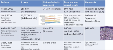
- Differentiating melanocytic Nevi from melanomas using HE stains (Hekler):
- 80% of nevi and 82% of melanomas were correctly classified.
- It is remarkable that only HE was used, as in normal histopathology other markers and stains are also used by the pathologist
- Lesion classification (Lenni):
- These slides were obtained from three different labs, which is a very challenging work because qualitative staining is always slightly different.
- Even so, they were able to have a very good results, for instance, 78% of the cases were correctly classified and only a few lesions were unclassifiable.
- Classification categories were divided into melanocytic, squamous, basaloid and “other lesions”.
- Detecting specific features of skin cancers and melanocytes nevi (Kucharski):
- The authors tried to find some nest of melanocyte to detect and delineate (segment) and to detect these lesions. And they did it with very good specificity ((exclusion when absent, precision) and quite reasonable sensitivity (confirm when present, recall).
- They tried to detect basal cell carcinoma (BCC), dermal nevi and seborrheic keratosis, the latter with with precision and recall.
Marcial Garcia-Rojo, MD. Teledermatopathology. 8th World Congress of Teledermatology, Skin Imaging and AI in Skin diseases – November 2020