Dermoscopy Imaging in Teledermatology
- With dermoscopy, things get simpler, fortunately. Dermoscopy is so important for us to make a diagnosis, especially for those of us that work in oncology.
- Sometimes even when the clinical image is bad, we can make a diagnosis because we do get a good dermoscopic image.
- As an anecdote by the presenter: “I always tell them as a joke that you need to take a course to take a good clinical picture, but you need to take a course to take a bad dermoscopic image.
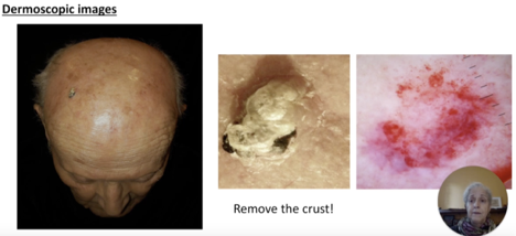
- Indeed everything is set to capture the image: the distance is the same, the lighting is the same, the colors are preserved…the most common mistake then with dermoscopy is to take pictures with the crust: it needs to be removed so that you can see what’s below the crust.
- Also, when the lesion is big, it’s important to have a closeup image and then one farther away. Nowadays, basically all dermatoscopes include a scale.
- Please also refer to the articles imaging in dermoscopy of inflammatory lesions (Dr Tejasvi) and pigmented lesions.
Paola Pasquali, MD. Digital Photography for Teledermatology Systems and Devices. 8th World Congress of Teledermatology, Skin Imaging and AI in Skin diseases – November 2020